Description
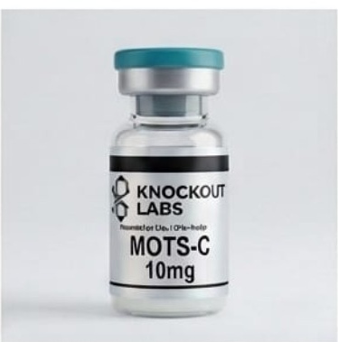
MOTS-c identity and the 10 mg listing
MOTS-c takes its name from a short open reading frame in mitochondrial 12S ribosomal RNA. The published human reference is a 16-residue peptide. “Mitochondrial-derived” describes its biological origin in the research literature. The name alone does not identify how the commercial material was produced. [M1]
| Check | What to compare |
|---|---|
| Peptide sequence | The human sequence used by Bhullar and colleagues was MRWQEMGYIFYPRKLR. Check the sequence and both termini on the specification. [M6] |
| Listed quantity | 10 mg is a nominal mass. Check the content basis and whether the amount includes a salt, water or other material. |
| Solubility and handling | Request data for the supplied form. A solvent used in a paper may suit a different preparation or assay. |
The MOTS-c compound profile gives the wider sequence and naming context.
Matching the analytical record to the lot
Ask technical support which lot is being offered and request the available MOTS-c documents. Check that the identifier, reported mass and method match the specification. A purity chromatogram alone cannot tell you how much peptide is in the container. See the testing methods guide and batch-report information.
Keep the container label and lot report together. For solution-based work, record the declared content basis, final volume and solvent. Follow the storage conditions supported for the actual preparation. The storage and stability guide explains why unopened material and a prepared solution need separate evidence.
Ordering and technical information
The purchase section shows current price and availability. Knockout Labs supplies UK purchasers aged 18 or over, with manual order review before acceptance. Products are Research Use Only. Not for human or veterinary use. Read the buyer-eligibility policy and delivery information, or contact technical support for the applicable specification and lot documentation.
MOTS-c research in mice and people
In exercise experiments, researchers administered MOTS-c to mice and measured physical capacity. In the human studies they measured the body’s own MOTS-c during or after exercise. Sample type and timing differed, and the human observations do not establish an effect from taking this product. [M3] [M4] [M5]
Read the metabolic, exercise and cell studies
Why MOTS-c is researched
The central question is how a small peptide can connect changes in cellular metabolism with a broader adaptive response. Researchers have examined glucose handling, stress-related gene expression and skeletal-muscle function. Those questions involve several distinct levels of evidence: biochemical changes inside cells, results from administering peptide to animals, and measurements of naturally occurring MOTS-c in people.
Metabolism and AMPK signalling
Lee and colleagues’ original experiments connected MOTS-c with changes in folate-linked and purine-synthesis pathways and activation of AMPK. AMPK is an energy-sensitive protein kinase: it can alter the activity of other proteins by phosphorylation. The authors also reported that treatment prevented insulin-resistance phenotypes associated with ageing or a high-fat diet in their mouse experiments, and reduced diet-induced obesity. [M1]
This places MOTS-c in a metabolic-regulation research programme, but an AMPK-related result is not a complete account of its mechanism. Changes in an upstream metabolic pathway, AMPK activation and whole-animal glucose handling are different observations. Their relationship should be examined with the controls used in the individual experiment, rather than treating the name of a pathway as proof of a universal effect.
Movement into the nucleus under metabolic stress
Kim and colleagues reported in 2018 that MOTS-c moved into the nucleus during metabolic stress in an AMPK-dependent manner. Under glucose restriction, it was associated with changes in nuclear gene expression, including genes with antioxidant response elements. The study also reported interactions with stress-responsive transcription factors such as NRF2. [M2]
A transcription factor helps regulate which genes are expressed; an antioxidant response element is a regulatory DNA sequence. This work therefore asks how a metabolic signal reaches a gene-expression programme. It supports investigating stress-dependent localisation and transcriptional responses, rather than assuming that the peptide remains in one cellular compartment or acts identically in unstressed and stressed cells. [M2]
Muscle bioenergetics: a more recent mechanistic result
Gudiksen and colleagues’ 2026 paper used two transgenic mouse models. The mitochondrial response to administered MOTS-c depended on AMPK and the metabolic regulator PGC-1α. They reported altered mitochondrial function without an apparent increase in respiratory-protein content, alongside lower reactive-oxygen-species emission. [M5]
This distinguishes a functional change from simply measuring more mitochondrial proteins. The findings concern the experimental mouse models. [M5]
Exercise findings: what was administered and what was measured?
Reynolds and colleagues’ 2021 study included both mouse experiments and a human exercise component. Administered MOTS-c improved measured physical capacity in young, middle-aged and old mice. In the human component, investigators measured endogenous MOTS-c following exercise and reported increases in muscle and circulation. The people in that exercise experiment were not given MOTS-c. [M3]
This distinction prevents an observational human result being described as a successful peptide-administration trial. A rise after exercise may help identify an adaptive signal; it does not establish what happens when an external preparation is administered to a person.
The human exercise literature is also not uniform. Ramanjaneya and colleagues studied women with polycystic ovary syndrome and healthy controls. Eight weeks of moderate exercise training did not change circulating MOTS-c levels in that study, although the experiments found responses to lipid and insulin challenges. An acute post-exercise measurement and a resting measurement after a training programme ask different questions. [M4]
In Gudiksen’s human one-legged exercise experiment, interstitial MOTS-c increased without a corresponding change in the arterial–venous difference across the leg. The investigators therefore questioned whether skeletal muscle supplied the circulating MOTS-c in that setting. [M5]
| Evidence layer | What the cited work contributes | What to keep separate |
|---|---|---|
| Cellular metabolism [M1] | Pathway and AMPK-related observations | A cellular response versus a whole-organism outcome |
| Nuclear signalling [M2] | Stress-dependent localisation and gene expression | Peptide location, transcription and functional response |
| Administered peptide in mice [M3] [M5] | Physical-capacity and mitochondrial-function results | Species, model, preparation and endpoint |
| Human exercise observations [M3] [M4] [M5] | Endogenous measurements with differing results | Observation after exercise versus administering MOTS-c |
Choosing useful laboratory endpoints
A study of metabolic signalling benefits from more than a single positive marker. For example, if the question concerns glucose handling, a transporter-localisation result, glucose uptake and a change in cell number should be distinguished. Bhullar and colleagues investigated mitochondrial fusion and GLUT4 movement in U-2 OS cells, a human osteosarcoma-derived cell line, linking mitochondrial organisation to the transporter’s response. That is a more specific experimental question than whether MOTS-c broadly “improves metabolism.” [M6]
Similarly, a nuclear-localisation experiment should document the stress condition and how nuclear and non-nuclear material were distinguished. A circulating-peptide measurement needs its sample timing and assay method. These are practical reading questions for the studies, not an instruction to reproduce their administration protocols.
When comparing papers, record the peptide sequence and modifications as well as the biological model. A named analogue, a labelled experimental peptide and an unmodified reference sequence should not be treated as identical reagents solely because they appear in the same research field. The exact supplied form is therefore part of experimental interpretation, not just catalogue administration.
Product questions
Does human exercise research show that administered MOTS-c works in people?
No. The human studies discussed here measured endogenous MOTS-c in an exercise or metabolic setting. Their results should not be relabelled as evidence from administering this product.
Does the 10 mg listing define a solution concentration?
No. A listed mass is not a concentration. A laboratory solution record needs a defined content basis and a measured final volume, as well as a compatible preparation method.
Is every MOTS-c preparation equivalent?
No. Check sequence, terminal modifications, counterion, content and formulation. Published experiments and the supplied lot need to be compared on those details.
Selected research sources
- M1. Lee et al. (2015). MOTS-c and metabolic homeostasis. Cell Metabolism. DOI: 10.1016/j.cmet.2015.02.009.
- M2. Kim et al. (2018). MOTS-c nuclear translocation under metabolic stress. Cell Metabolism. DOI: 10.1016/j.cmet.2018.06.008.
- M3. Reynolds et al. (2021). MOTS-c, exercise and muscle homeostasis. Nature Communications. DOI: 10.1038/s41467-020-20790-0.
- M4. Ramanjaneya et al. (2019). Lipids, insulin and circulating MOTS-c. Clinical Endocrinology. DOI: 10.1111/cen.14007.
- M5. Gudiksen et al. (2026). MOTS-c and muscle mitochondrial bioenergetics. Free Radical Biology and Medicine. DOI: 10.1016/j.freeradbiomed.2026.01.002.
- M6. Bhullar et al. (2021). Mitofusion and MOTS-c-induced GLUT4 translocation. Scientific Reports. DOI: 10.1038/s41598-021-93735-2.