Description
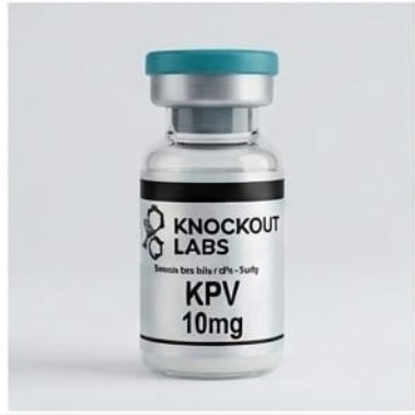
KPV 10 mg and the supplied peptide form
KPV stands for lysine–proline–valine, the last three residues of α-melanocyte-stimulating hormone (α-MSH). A paper that uses the parent hormone or a modified KPV preparation describes a different experimental material. [K0] [K2]
| Detail | Why it matters for this listing |
|---|---|
| Terminal groups | Check whether the carboxyl end is a free acid or an amide. The three-letter sequence alone leaves that distinction open. |
| 10 mg listed amount | Ask whether the figure represents peptide content or total material including a counterion or water. Working concentration also depends on the final volume. |
| Identity and content | Match the declared form and lot identifier to the analytical report. A chromatographic peak-area percentage cannot establish the number of milligrams in the container. |
The KPV compound profile covers reference identities. ChEBI reports an average mass of 342.440 Da for its free-acid entry. Use that value for a lot calculation only if the specification matches that form. [K0]
Checking the KPV lot before ordering
Request the specification and available KPV lot documents. Ask which terminal form is supplied, what the nominal fill includes and which method supports any quantity claim. The batch-report information page explains how the records are organised.
For a short tripeptide, check that the chromatographic method separates the intended analyte from related components. The analytical testing guide explains how identity, relative peak area and measured content answer different questions.
On receipt, match the container’s lot number to the report. Retain the handling instructions for that form and container alongside the specification. The storage and stability guide explains what evidence to look for.
UK eligibility and availability
The product panel shows the current price and stock. Knockout Labs supplies UK customers aged 18 or over and reviews orders before acceptance. Read the research-use and buyer eligibility policy and UK delivery information.
Research Use Only. Not for human or veterinary use.
What has KPV research measured?
Researchers have measured KPV transport into cells, changes in inflammatory markers in mice and the effects of a nanoparticle carrier. These studies used defined experimental preparations. They do not establish an effect in people or a property of the lot offered here. [K1] [K2] [K3]
Read the transport, formulation and antimicrobial studies
Why KPV is researched
The research asks several connected questions: can cells take up this short peptide, does it alter a measured inflammatory response, and does a delivery system change where the peptide reaches? Each needs a different experiment. Uptake establishes access to a cell; a signalling assay examines a response; a formulation study tests the peptide together with its carrier.
Cell uptake through PepT1
Dalmasso and colleagues studied human intestinal epithelial cell lines and Jurkat T cells, alongside two induced mouse-colitis models. They used uptake experiments to investigate PepT1, a transporter for short peptides, and measured NF-κB and MAP-kinase signalling and cytokine output. KPV entered cells through PepT1 and reduced the stimulated signalling and inflammatory-cytokine responses under the tested conditions. [K1]
The mouse experiments also reported lower inflammatory-cytokine expression in DSS- and TNBS-induced colitis. The work links cellular access with downstream measurements, rather than establishing that KPV simply blocks every inflammatory pathway. PepT1 is a transporter, not a demonstrated universal KPV receptor. Differences in transporter expression are therefore relevant when comparing cell models. These experiments do not establish a clinical response to this product. [K1]
What the melanocortin-receptor comparison showed
Kannengiesser and colleagues tested KPV in DSS colitis and a T-cell-transfer model in mice. They reported improved recovery of body weight, reduced inflammatory infiltrates in tissue and lower colonic myeloperoxidase activity. Myeloperoxidase is an enzyme used here as an indicator associated with inflammatory-cell activity. [K2]
The researchers also tested mice with a nonfunctional melanocortin-1 receptor. Effects persisted in that setting, supporting their conclusion that the response was at least partly independent of MC1R signalling. This helps distinguish KPV from an assumption based solely on its α-MSH ancestry. It does not exclude every melanocortin-related mechanism or demonstrate receptor independence in every tissue. [K2]
A delivery system is part of the intervention
Xiao and colleagues loaded KPV into polymeric nanoparticles functionalised with hyaluronic acid, then incorporated the particles into a chitosan/alginate hydrogel. Their 2017 study examined delivery to colonic epithelial cells and macrophages and tested the system in a mouse-colitis model. The functionalised formulation reduced mucosal damage and TNF-α more effectively than the comparison KPV nanoparticle/hydrogel system. [K3]
The comparison concerns two delivery systems containing KPV. It is not evidence that an unformulated 10 mg container has the same distribution, persistence or biological effect. Carrier composition, particle properties and release behaviour are part of what was tested. When using this literature to frame laboratory work, record the full intervention rather than citing the peptide name alone. [K3]
Antimicrobial findings and their limits
Cutuli and colleagues’ 2000 laboratory study examined α-MSH peptides, including KPV, against Staphylococcus aureus and Candida albicans. They reported reduced bacterial colony formation and changes in yeast viability and germ-tube formation. The paper also investigated cAMP, a cellular messenger, as a possible contributor to the observed effects. These were laboratory endpoints, not treatment outcomes in people. [K4]
A later published comment by Rauch and colleagues reported difficulty reproducing strong anti-Candida effects in their melanocortin-peptide assays. The disagreement makes the exact peptide, organism strain and assay conditions essential to interpreting this literature. It should not be collapsed into either a universal antimicrobial claim or a definitive negative result for the offered KPV lot. [K5]
The selected studies provide preclinical questions for investigation. They do not establish KPV as a treatment for infection or intestinal disease, and they are not a clinical-use protocol for this research material. This is a focused reading of relevant papers, not an exhaustive survey of every KPV study.
Turning the literature into a clear laboratory question
An experiment is easier to interpret when the input, model and endpoint are stated separately. For example, reduced cytokine output could reflect altered signalling, fewer viable cells or a difference in peptide access. Measuring viability and the intended response helps distinguish those possibilities.
| Research question | What should be distinguishable |
|---|---|
| Is the peptide entering the cells? | Uptake from a downstream biological response |
| Has a stimulated response changed? | Signalling or cytokine output from changes in viable cell number |
| Does a carrier improve delivery? | Peptide, empty-carrier and formulated-peptide effects |
| Can two papers be compared? | Exact peptide form, cell type, stimulus, timing and analytical endpoint |
This is an interpretation framework, not a validated protocol for the offered lot. A positive result in one system should be reported with that system’s conditions. A negative result also needs the material identity and method stated before concluding that it contradicts a different model.
Product questions
Is KPV the same as α-MSH?
No. KPV represents a three-residue sequence from the larger peptide. The parent molecule’s findings cannot all be assigned to the fragment, and the exact tested form still matters.
Is KPV interchangeable with BPC-157?
No. They have different sequences and molecular sizes. Equal milligram quantities do not establish equal molar quantities or comparable biological activity. The KPV and BPC-157 comparison explains the identity distinction.
Does the nanoparticle paper describe this product’s formulation?
No. This listing does not establish that the material contains that carrier system. Formulation properties must be supported for the actual supplied item.
Can I use a purity percentage to calculate how much KPV was supplied?
A relative peak-area percentage describes what the method detected. To establish peptide mass, ask for a quantitative result, the unit reported and the lot number to which it applies.
Selected research sources
- K0. ChEBI. Lys-Pro-Val, CHEBI:160254.
- K1. Dalmasso et al. (2008). PepT1-mediated KPV uptake and inflammatory responses. Gastroenterology. DOI: 10.1053/j.gastro.2007.10.026.
- K2. Kannengiesser et al. (2008). KPV in two mouse-colitis models. Inflammatory Bowel Diseases. DOI: 10.1002/ibd.20334.
- K3. Xiao et al. (2017). KPV nanoparticle delivery in experimental colitis. Molecular Therapy. DOI: 10.1016/j.ymthe.2016.11.020.
- K4. Cutuli et al. (2000). Antimicrobial experiments with α-MSH peptides. Journal of Leukocyte Biology. DOI: 10.1002/jlb.67.2.233.
- K5. Rauch et al. (2009). Comment on anti-Candida peptide findings. Journal of Leukocyte Biology. DOI: 10.1189/jlb.1008614.